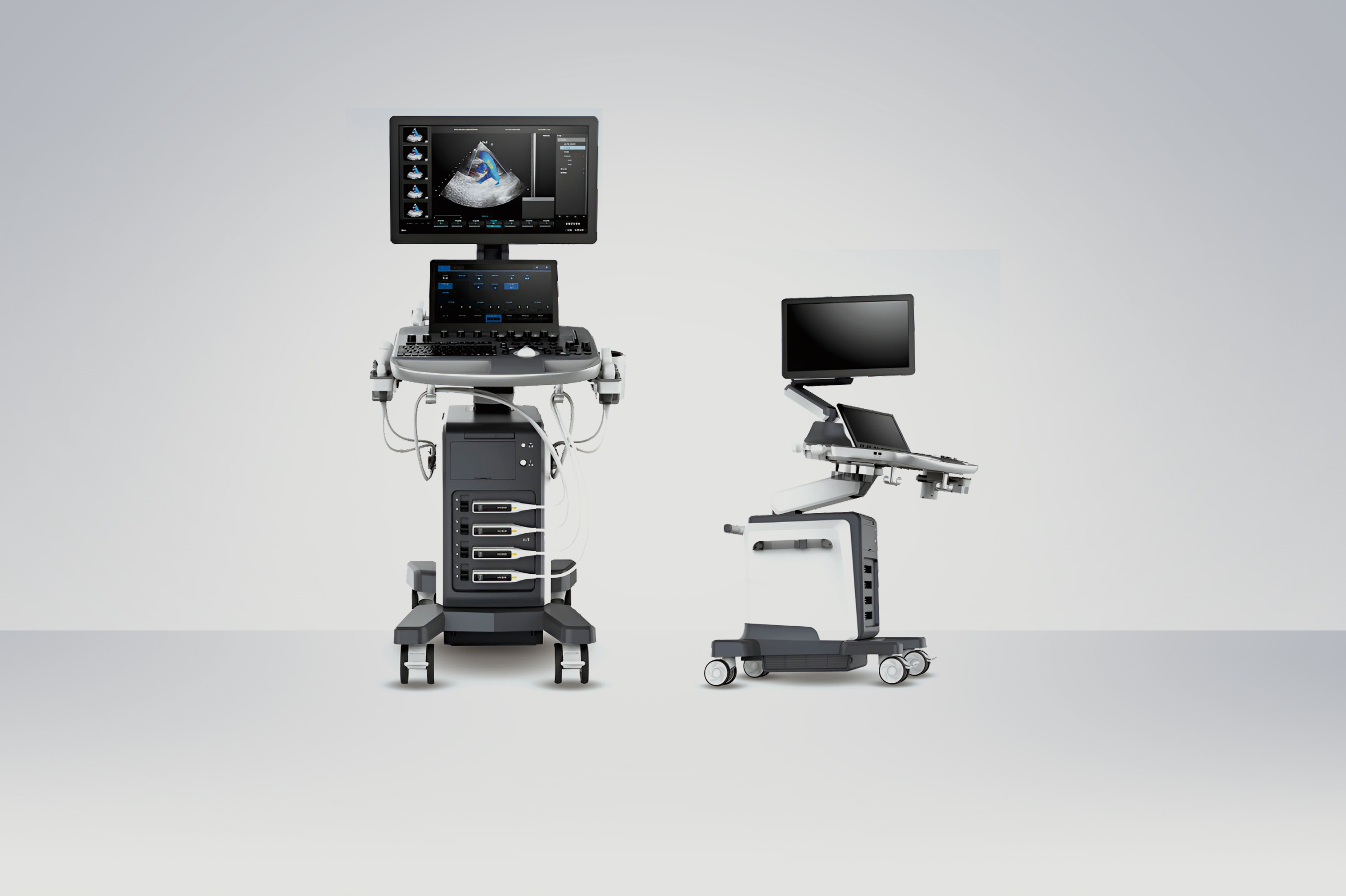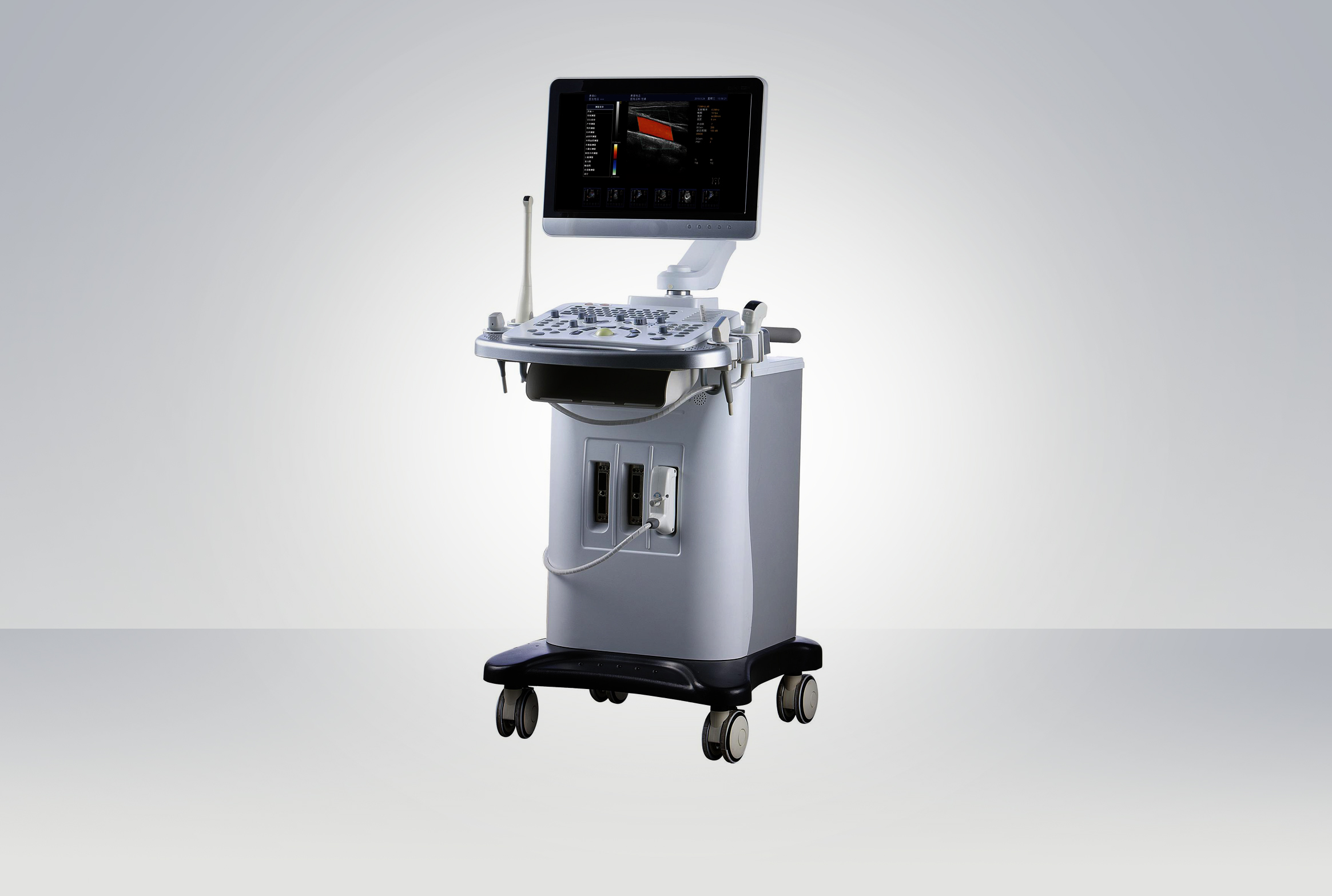

TBC-9760 Color Doppler Ultrasonic Scanner
| Item No.: | TBC-9760 |
| Min. Order: | 1 Piece |
|---|
General Application:
Abdomen, OB/Gynecology, Vascular, Cardiology, Small Parts, Breast, Urology, Pediatrics, TCD, Intra operative, and Musculoskeletal
Main Technical Parameter:
Check mode: abdomen, cardiac, gynaecology, obstetrical, kidney, Urology, blood vessel, small organ;
Probe type: Convex array probe, Linear array probe, phased array probe, Transvaginal probe, Micro-convex probe, 4D volume probe;
Imaging mode: B mode,2B mode,4B mode, M mode, Anatomy M-mode, Curve anatomy M mode, Color CM mode, Color mode, PDI mode, DPDI mode, CW mode, PW mode, Panoramic imaging, Elastic imaging, Contrast Imaging, TDI imaging, 3D mode, 4D mode;
Transducer ports: 4 fully activated transducer ports;
With built-in gel warmer;
Digital Video Recorder, record the screen and audio information, save it into hard disk;
Size and weight: W962mm,D591mm,H1348~1751mm, Weight: about 100kg;
Power supply: 100V-240VAC, 50Hz/60Hz, Battery specification: 21.6V/17Ah;
Monitor: 23.8-inch high-resolution color LED monitor;
Resolution: 1920×1080;
The display screen rotates independently: ± 90°;
Display screen folded up and down: 90°;
Touch screen: 13.3-inch touch screen; Resolution: 1920×1080;
Touch screen Angle is adjustable 0~55°;
Support operate with glove;
The Control Panel: User-centered design for easy operation;
Panel button backlight design: 0-9 level adjustable;
10 user-defined keys: Q1-Q8, Save1/Save2;
8-segment TGC slide, backlight design;
Standard text keyboard;
The key tone adjustable;
Trackball sensitivity adjustable;
Dedicated palm placement area to reduce wrist fatigue injury;
Independent lifting of the control panel: 0-220mm;
Control panel swivel of -35 degrees to +35 degrees;
Synchronize main screen display: Real-time remote image viewing thru third party software;
Application must be downloadable for Android;
Wireless image/video transmission to a smart device;
B Mode scan depth is at least 40cm. With adjustable frequency;
Auto optimization for Gain, Dynamic range and TGC.
Maximum frame rate: At least 800 f/s
Tissue Harmonic Imaging/ Pulse Inversion Harmonic Imaging
M mode and with different display format
Display: At least 3 sample lines simultaneously.
The angle and position of sample lines can be adjustable.
Color Flow Mapping Mode
PDI/ DPDI
PW sample volume is 0.5-40mm
CW with a velocity range of 0-45m/s
Tissue Doppler Imaging
Compound Imaging
Tissue Specific index
High Resolution Flow
High Pulse Repetition Frequency
Simultaneous Mode (Triplex):
a. AUTO IMT
b. AUTO NT
c. AUTO EF
d. AUTO BLADDER
Automatic Measurement of BPD / HC / AC / FL / HL, Meansurement by hands for Cereb / Vp / CTAR / CTDR / Cardiac axis / PL / AFI
Biopsy Guide
Real-Time 2D
Color Panoramic Imaging
Vis needle or equivalent
3D/4D Module (Application)
a. Static 3D/4D and Realtime 4D
b. Color 3D/4D image
c. Presets/ user defined parameters for 3D/4D applications
Freehand 3D
Strain Elastography with Strain Ratio Measurement
Biopsy Guide
Customized Workflow Protocol for Vascular and Obstetrics Applications
DICOM 3.0:
Store/C Store/ Worklist / MPPS/Print/ SR/Q&R
OPERATING SYSTEM: LINUX Internal storage: At least 512GB SSD and 1TB HDD
2D cine storage time setting:
a. Retrospective storage
b. Prospective storage
c. Freeze Storage
4D cine storage time setting
a. Retrospective storage
b. Prospective storage
Directly Store to USB Drive
Export data to USB drive or DVD
Image format: BMP, JPG, TIF
Cine format: AVI, MP4
Report format: PDF, TXT, HTML, RTF
DICOM format
Post processing and post measurement
Backstage storage: quick switch of DICOM cine
USB port 2.0:4
USB 3.0: 2
Video In and Output Ports
VIDEO/S-VIDEO (TV-NTSC, TV-PAL)
Multi frequency imaging capability for optimal resolution and penetration.
Single Crystal Convex Probe
a. Applications: Abdomen, Obstetrics
b. Bandwidth range: 1-7MHz
c. Field of View: at least 60°
d. Convex radius: at least 45mm
e. Depth of at least 40cm
Endocavity Convex Probe
a. Applications: Gynecology, Urology
b. Bandwidth range: 2-14 MHz
c. Field of View: 180º
d. Convex radius: at least 10mm
e. Depth of at least 15cm
Linear Array Probe
a. Applications: Peripheral Vascular, Superficial and small parts.
b. Bandwidth range: 2-16MHz
c. Width of View: at least 45mm
d. Depth of at least 10cm
Volume Probe (3D/4D)
a. Application: Abdomen, Obstetrics, Gynecology
b. Bandwidth range: 1-7MHz
c. Field of View: at least 65º
d. Curvature radius: at least 40mm
e. Depth of at least 30cm
ACCESSORIES
1.Built-in battery
2.Ground Lead
3.AC Power Cord
4.Basic User Manual: English
5.Basic Service Manual: English
6.English – Quick Start Guide
7.B/W Thermal Printer